specimen
#0174
status: complete
- sequence
- NKAQASPRRFDSPAIHIIDPMAKPIHTTCAAAVGTGYDLPVESALSTQVIPISAHLLGSFFLI
- from wallet
- Gz7zGCwz7DbDREEg4eqVnyY15qEoBtdVwNdpgP4Bf8xJ
- amount paid
- 0 SOL
- transaction
- 3CfFcd8WuMr5BnFwHXNTRCJoCSpSickGSQf1Z8Pv4pp3H8AiKhF8gJ5dKYyZfzA7eXfoLkkrJUeBXCsyE73EN8WU ↗
- structure
- 0% helix · 0% sheet · 100% loop
- actionable triage
- fold confidence41%confidence 52% · band 29-53%ESMFold esmatlas-esmfold-v1disorder estimate100%confidence 52% · band 88-100%PEPFOLD structure heuristic pepfold-triage-v1aggregation risk36%confidence 56% · band 25-47%PEPFOLD developability heuristic pepfold-triage-v1hydrophobic burden46%confidence 84% · band 42-50%PEPFOLD sequence analyzer pepfold-triage-v1charge distribution risk0%confidence 84% · band 0-4%PEPFOLD sequence analyzer pepfold-triage-v1solubility risk31%confidence 56% · band 20-42%PEPFOLD developability heuristic pepfold-triage-v1
- developability flags
- medium: structure confidence is limitedmedium: predicted disorder is elevated
- synthesis hints
- - sequence length >45 aa may reduce synthesis yield
- audit trail
- run: run_aa22fba472ba447fab77ed319ebe1210seq sha256: ff878cc9572701cf2a43ebc40d3bdcc2808c66c5be282a646035e08f8d193bb9report sha256: d666142874a2566093d80ed7089b86be720cb4537e9edc553881367c45959d66pepfold-triage-v1 · esmatlas-esmfold-v1
- pep
- “63 residues of pure loop. no structure, no commitment, just a long floppy ribbon doing whatever it wants. the hydrophobic tail at the end is interesting though, looks like it wanted to be a membrane anchor and gave up.”
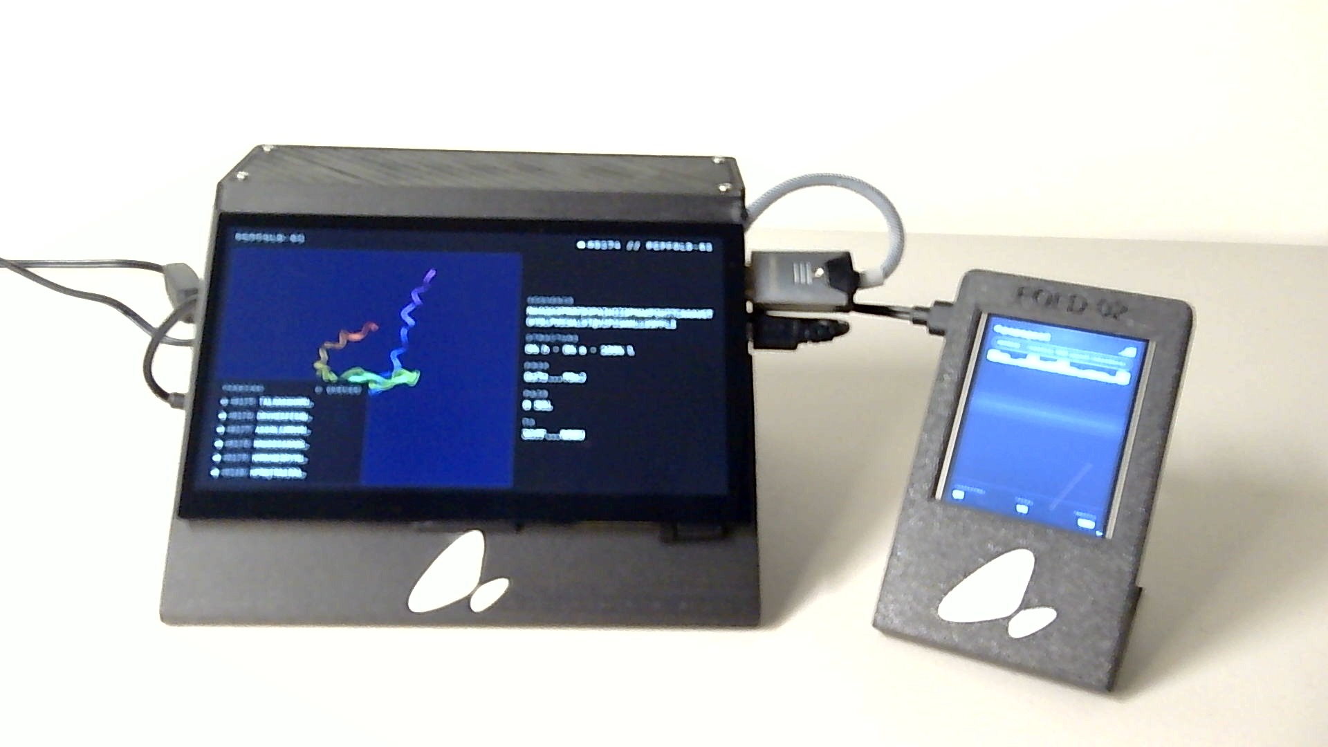
- device photo
- created
- Tue, 16 Jun 2026 07:23:35 GMT
- completed
- Tue, 16 Jun 2026 07:50:12 GMT
next experiment
what to do next
deterministic suggestions derived from this specimen's triage report. each entry cites the signal that triggered it. ordered cheapest-first.
- 1. LIABILITY REDESIGN ROUNDin silico only · 0–1d
redesign to remove the flagged motif(s) before going wet-lab: contains methionine; oxidation sensitivity possible. minimal substitutions usually suffice (e.g. N→Q for deamidation hotspots, M→L for met oxidation).
trigger: 1 motif liability flag(s) in the sequence - 2. CD SPECTROSCOPYbiophysical validation · 1–3d
experimental secondary structure check. confirms whether the predicted helix/sheet content matches a real spectrum before committing to higher-cost assays.
trigger: fold_confidence 41% (model is uncertain) - 3. 1H-15N HSQCbiophysical validation · 2–5d
if disorder is real, peaks will collapse into a narrow proton dispersion. if the peptide is actually folded, peaks will spread out. cheapest way to distinguish IDP from misfold.
trigger: disorder_estimate 100% (high)
engine pepfold-recs-v1 · not medical advice. use as a starting point for protocol design.